Analyzing Western Blots, etc.
Not what you're looking for?
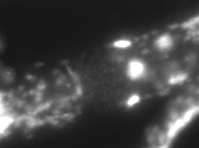
I took photographs of 3 stains: SDS-PAGE analysis of C. elegans protein extract (Coomassie Blue Staining), Western Blot of C. Elegans protein, and Indirect Immunofluorescence Staining. There are 3 photos attached .How do you analyze these stains? I need help discussing these results.
Purchase this Solution
Solution Summary
The expert analyzes Western Blots. The SDS-PAGE analysis of C. elegan protein extracts are examined.
Solution Preview
OTA ID# 101743
I took photographs of 3 stains: SDS-PAGE analysis of C. elegans protein extract (Coomassie Blue Staining), Western Blot of C. Elegans protein, and Indirect Immunofluorescence Staining. There are 3 photos attached .How do you analyze these stains? I need help discussing these results.
This is truly and exiting problem!
Lets start by understanding what each image means and then I will tell you the information you can get off the image.
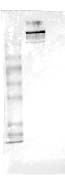
The SDS-PAGE is a technique to separate proteins by their size (mass). PAGE stands for polyacrylamide gel electrophersis, this material basically serves as a matrix or mesh and proteins will migrate through its holes. A small protein will fit through the holes better than a large one and will migrate faster. SDS stands for sodium dodecyl sulfate, and is a anionic detergent, it essentially disrupts all noncovalent interactions in your protein preparation. The Coomassie blue stain is merely to visualize the protein you have separated using this technique.
On your gel image you have 5 lanes. I will tell you what I think each lane contains since you have not specified it.
From left to right:
1- This is the protein marker. Each band has a specified mass. This serves to you as a reference so that you can estimate the mass of protein bands that have migrated a similar distance. It also serves a very important purpose besides telling about size of the proteins. Imagine protein purification did not work and you did not add protein markers to your SDS-PAGE. After staining the gel you would still see nothing because ther is no protein. How would you know that your Coomassie blue stain worked? Well, the protein markers are usually purchased from companies, so it is "guaranteed" that they have protein. So the fact that you see the bands is your gel is a good indicator that the staining method went well. Finally, the separation of the bands tells you that ...
Purchase this Solution
Free BrainMass Quizzes
How Well Do You Know Your Body?
This quiz will assess the different systems of the human body. It will examine everything from the organs to the cellular processes that occur.
Pregnancy Knowledge
How much do you know about being pregnant? Test your Pregnancy IQ with this quiz!
Do You Know Your Macromolecules?
This quiz will assess your knowledge of the macromolecules that are important to living things.
Basic Immunology Quiz
Intro to immuno quiz. Covers the basics of immunology and recognition of foreign substances by the body.
Basic Concepts in Neuroscience
This quiz provides a review of the basic concepts in neuroscience.