As the control center of the entire body, the brain is constantly sending and receiving millions of signals at any given moment. These exchanges of information control our movements, our drives, the function of our organs and our thoughts. There are different types of obstructions that can interrupt this activity. It is vital to detect these intrusions in order to relieve possible symptoms and restore complete brain functioning. These can only be determined by examining the brain internally to discover what is causing the interruptions.
There are many invasive and noninvasive methods of measuring neural activity. These include the following:
 Optical techniques rely on methods that change optical or visual properties of neurons in response to neural events like action potentials or the release of transmitters.¹ There are several different optical methods that can be used. Voltage sensitive dyes (VSDs) become fluorescent in response to changes in voltages in neurons. This makes individual action potentials detectable.¹ Calcium imaging also relies on dyes that become fluorescent when they bind to the calcium present in an action potential.¹ Synapto-pHluorin relies on a fusion protein that becomes fluorescent when exposed to the higher pH of the synaptic cleft.¹
Single-unit recording introduces an electrode into the brain to detect electrical activity from the adjacent neurons.

Multielectrode recording is performed by bundling fine electrodes to record the activity of hundreds of neurons at once.
Functional magnetic resonance imaging (fMRIs) is a technique in which the changes in cerebral blood flow are detected and used to indicate the activity of larger scale brain regions.¹
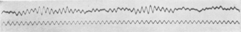
Electroencephalography (EEG) is a technique in which scalp electrodes monitor the average activity of neurons.
Magnetoencephalography (MEG) measures the functioning of the brain through measurement of electromagnetic activity.
References:
1. Methods of Monitoring the Brain. Retrieved from: http://www.macalester.edu/academics/psychology/whathap/UBNRP/Imaging/index.html